
Three-dimensional clarity for confident planning
Cone-beam computed tomography gives our surgeons a precise three-dimensional view of your jaw, nerves and sinuses — the foundation for safer, more predictable and truly personalised treatment.
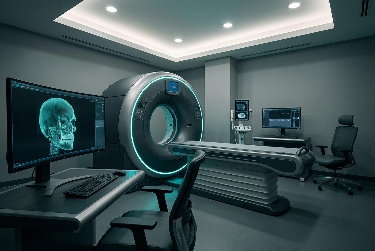 3D-Guided Surgical Planning
 3D cone-beam computed tomography, often shortened to CBCT, is an advanced imaging technology that produces highly detailed three-dimensional images of your teeth, jawbone, nerves, sinuses and surrounding structures. Unlike a conventional flat dental X-ray, a cone-beam scan allows our surgeons to view your anatomy from every angle, providing a level of clarity that is central to modern, precise surgical care.
This imaging forms the foundation of confident treatment planning across our practice. Whether you are considering dental implants, ridge expansion, the removal of complex wisdom teeth, or an assessment of nerve position, a cone-beam scan allows us to plan each step with accuracy and to anticipate and avoid potential complications.
Why three-dimensional imaging matters
Important structures such as nerves, blood vessels and the sinus cavities cannot be fully appreciated on a standard two-dimensional X-ray. By mapping these in three dimensions, we can measure bone with precision, identify the exact position of a nerve, and plan procedures such as implant placement using guided surgical techniques. The result is treatment that is safer, more predictable and tailored entirely to you.
- Precise planning for dental implants and guided surgery
- Accurate mapping of nerves to protect sensation during surgery
- Detailed assessment of bone volume, density and quality
- Evaluation of the sinuses, jaw joints and impacted teeth
- Clearer explanations, as you can see your own anatomy alongside us
A simple and comfortable experience
Having a cone-beam scan is quick, straightforward and entirely painless. You simply remain still for a short time while the scanner rotates gently around your head, typically capturing the images in under a minute. There is no discomfort, and the scan is performed using equipment selected to keep the radiation dose as low as reasonably achievable while still providing the detail your treatment requires.
Because the images are available immediately, we can review them with you during your visit, pointing out the relevant anatomy on screen and explaining clearly how it informs your care. Many patients find that seeing their own scan helps them understand their treatment and feel reassured about the plan ahead.
If you would like to understand your options with the benefit of precise, modern diagnostics, we invite you to arrange a consultation. Our team will explain how 3D imaging supports safer, more personalised care from the very first step.
3D Cone Beam Imaging — your questions
Cone-beam imaging uses a low dose of radiation, and we always follow the principle of keeping exposure as low as reasonably achievable. The detailed information it provides significantly improves the safety and precision of your surgical planning, and we are happy to discuss any concerns.
Not at all. The scan itself is usually completed in well under a minute, during which you simply sit or stand still while the scanner rotates gently around your head. The whole appointment is quick, comfortable and entirely painless.
A standard X-ray provides only a flat, two-dimensional image, which cannot fully show the position of nerves, the volume of bone, or the relationship between structures. A 3D scan reveals this detail, allowing far more precise and safer surgical planning.
Discuss 3d cone beam imaging with our team
Specialist assessment, 3D imaging and a calm, clearly-explained plan — arrange a consultation with the team at Maxilla Surgical Arts.